RUSSIAN JOURNAL OF EARTH SCIENCES, VOL. 16, ES3003, doi:10.2205/2016ES000573, 2016
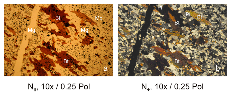
Figure 3. Two representative images illustrating the same thin sections segment of the Outokumpu 1844 biotite gneiss obtained in parallel (a) and cross (b) Nicols. Besides plagioclase feldspar (Pl), quartz (Qz) and magnetite (Mg), both images illustrate the presence of strongly deformed, almost acicular biotite crystals (Bi).

![]()
Citation: Duliu Octavian G., Tatiana I. Ivankina, Edward Herman, Calin Ricman, Ion Tiseanu (2016), Orientation distribution function of biotite platelets based on optical, thin sections and $\mu$-CT image analysis in an Outokumpu (Finland) biotite gneiss: Comparison with neutron diffraction texture analysis, Russ. J. Earth Sci., 16, ES3003, doi:10.2205/2016ES000573.
Copyright 2016 by the Geophysical Center RAS.
Generated from LaTeX source by ELXpaper, v.1.5 software package.